X-ray imaging is an important technique for a variety of applications including medical imaging, industrial inspection and airport security. An X-ray image shows a two-dimensional projection of a three-dimensional body. The original 3D information can be recovered if multiple images are given of the same object from different viewpoints. The process of recovering 3D information from a set of 2D X-ray projections is called Computed Tomography (CT).
Traditional CT scanners are based on a single moving source, rotating around the object to reconstruct. In this set-up, images are taken sequentially and the resulting reconstruction problem gives rise to a linear system of equations.
The innovation of X-ray emitter arrays allows for a novel type of X-ray scanning device with faster image acquisition due to multiple simultaneously emitting sources. Acquisition speed is an important factor in medical imaging because it can help to avoid artefacts from motion of the analysed tissue. Another major advantage of emitter arrays is that they result in light weight, highly mobile 3D imaging systems that can be taken to the patient rather than having to move the patient to a radiology suite.
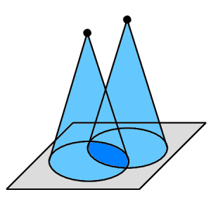
However, two or more sources emitting simultaneously can yield measurements from spatially and temporally overlapping rays. This imposes a new type of image reconstruction problem based on nonlinear constraints that traditional linear image reconstruction methods cannot cope with.
Oxford Mathematicians Raphael Hauser and Maria Klodt have derived a mathematical model for this new type of image reconstruction problem, and developed a reconstruction method that allows the recovery of images from measurements with overlapping rays. Based on compressed sensing, the method allows for reconstruction from undersampled data, which means that the number of reconstructed densities is higher than the number of measurements, which enables reduced doses (see the full paper).
The method has been successfully applied to real X-ray measurements in cooperation with Gil Travish and Paul Betteridge from industrial partner Adaptix, an Oxford-based start-up company. The Adaptix scanners acquire images with a flat panel of comparatively small emitters with small opening angles of the emitter cones, arranged in a fixed grid which can allow for small devices with reduced doses.
The new image reconstruction method opens new possibilities for X-ray scanner design, because it allows for a new class of hand-held X-ray scanning devices, where emitter and detector positions cannot be aligned exactly, and overlapping of emitter cones cannot be avoided.