Mathematical modelling played a key role in describing the spread of the COVID-19 pandemic; now a different kind of maths is helping us understand how immune cells interact in the lungs of patients with severe COVID-19.
In a damaged lung with a massive immune cell infiltrate, as seen with severe COVID-19 infection, it can be difficult to figure out which cells are involved in causing lung injury.
Understanding this provides a first step to identifying the cells or pathways that can be targeted therapeutically.
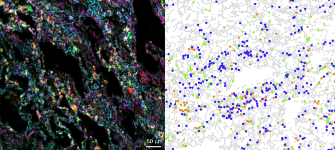
In a Nature Communications study Oxford Mathematicians Joshua Bull and Helen Byrne used a suite of mathematical tools to describe the spatial interactions between different cell types in complex images of COVID-19 lungs. The images, generated by Prof Ling-Pei Ho’s group in the Weatherall Institute for Molecular Medicine in Oxford, show the locations of 35 different cellular markers and provide a detailed map of the immune landscape of lung biopsies from patients with fatal COVID-19 infection.
"This work really highlights the benefits of multidisciplinary science” says Helen, “we came together as a new team, learned to speak each other’s languages and generated insight into lung disease that wouldn’t have been possible otherwise."
The authors used mathematical methods for describing spatial interactions between sets of points to identify associations between different cell types. This revealed a specific association between a highly inflammatory cluster of immature neutrophils, CD8 T-cells and regenerating lung stem cells in the most damaged areas of the lungs. Neutrophils and T-cells are both types of immune cells. Neutrophils are part of the innate immune system and are one of the first responders in the immune system. T-cells, on the other hand, are part of the adaptive immune system, providing a more specific response to infection than the neutrophils.
Circulating immature neutrophils are unusual, but are known to be increased in the blood of patients with severe COVID-19. This study provides the first evidence of their presence in the lungs of those with severe COVID-19, as well as their interaction with CD8 T-cells and lung stem cells. It highlights a potential role for immature neutrophils in lung injury and the possibility that this interferes with the ability of the lung to regenerate after severe viral infections. Crucially, without using the language of mathematics to identify spatial correlations between these cell types, the clusters would have remained unidentified.
The study was a collaboration between immunobiologists and computer scientists based at the Weatherall Institute for Molecular Medicine and mathematicians at the Mathematical Institute’s Wolfson Centre for Mathematical Biology. It demonstrates how mathematics combined with biological insight can be used to discover interactions and cellular networks which are otherwise impossible to identify.
Image (left): immune cells in a COVID-19 infected lung, showing groups of immune cells crowded around damaged airways (empty spaces)
Image (right): key cell types in COVID-19 lung tissue, represented as a spatially embedded network: nodes represent cell centres, and are coloured according to cell type, while nodes are connected by an edge if the cells are in direct physical contact.


